



Why Pregnancy Trimesters Are Divided: A Clear Medical Guide

Índice
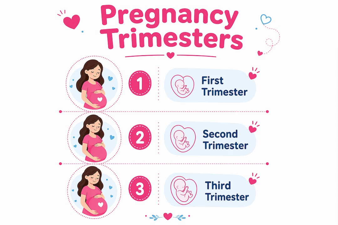
Pregnancy is divided into three trimesters to organize the 40-week gestation period into manageable stages, each reflecting distinct fetal development and maternal changes. Think of it as a clinical roadmap: rather than tracking 280 days as one continuous stretch, the trimester system groups weeks into three phases that align with key biological shifts and prenatal care milestones. Here is a quick overview of the timing:
- First trimester: weeks 1–13 (conception through early organ formation)
- Second trimester: weeks 14–26 (fetal growth, increased maternal energy)
- Third trimester: weeks 27–40 (fetal maturation, preparation for birth)
The division originated from the simple idea of splitting “nine months of pregnancy” into three equal parts. As you will see, the biology is more layered than that, and clinicians have developed richer frameworks to complement this traditional model.
Why the first trimester matters most biologically
The first trimester runs from conception through the early teens of pregnancy, and it carries the highest biological stakes of the entire pregnancy. This is when the embryonic period unfolds: rapid cell differentiation produces the neural tube, heart, limbs, and all major organ systems in a matter of weeks. By week 8, the embryo has transitioned into a fetus with recognizable human features.

Your body is working overtime during this phase. Hormonal surges, particularly in human chorionic gonadotropin (hCG) and progesterone, trigger the symptoms most people associate with early pregnancy: nausea, fatigue, breast tenderness, and frequent urination. These changes affect nearly every organ system, often before you even know you are pregnant.
The first trimester is also the period of greatest sensitivity to teratogens, substances or exposures that can disrupt fetal development, especially during early weeks when organ formation happens. Alcohol, certain medications, and infections like rubella pose the highest risk during organogenesis. That is why early prenatal care focuses so heavily on this window.
Clinical milestones in the first trimester:
- Confirmation of pregnancy via blood or urine hCG test
- First prenatal visit, typically between weeks 8 and 10
- Early ultrasound to confirm gestational age and viability
- Blood tests screening for genetic conditions, blood type, and infections
- Optional nuchal translucency ultrasound around week 12 to assess chromosomal risk
Pro Tip: If you are experiencing severe nausea or vomiting, talk to your provider early. Hyperemesis gravidarum affects a smaller subset of pregnant people and can require medical treatment to prevent dehydration.
What changes during the second trimester
The second trimester spans weeks 13–27, and for most people, it brings welcome relief. The intense nausea of the first trimester typically fades, energy returns, and the pregnancy becomes visibly apparent as the belly grows. Fetal development shifts from organ formation to organ maturation and rapid size increase.
By week 18, your baby’s movements, called quickening, become perceptible. The fetus develops hearing, swallowing reflexes, and a layer of vernix caseosa (a protective coating on the skin). Organ systems that formed in the first trimester now refine their function. You can track these changes week by week, including what is happening around week 18.

Maternal physiology keeps evolving too. Blood volume increases substantially, the uterus rises above the pelvis, and round ligament pain becomes common as the body accommodates the growing fetus. Skin changes like the linea nigra and melasma often appear during this period.
Key second-trimester assessments:
- Anatomy scan ultrasound (typically weeks 18–20) to check fetal organ development
- Maternal serum screening (quad screen) for chromosomal and neural tube conditions
- Cervical length measurement if preterm birth risk is elevated
- Blood pressure monitoring for early signs of preeclampsia
Pro Tip: The anatomy scan is one of the most detailed ultrasounds of your pregnancy. Write down your questions beforehand so you can make the most of that appointment with your provider.
How the third trimester prepares you for birth
The third trimester covers weeks 28 through 40, and its biological focus shifts almost entirely to preparation for life outside the womb. The fetus gains the majority of its birth weight during this period, with fat deposits forming under the skin to regulate body temperature after delivery. Lung maturation, including the production of surfactant, is one of the most critical developments of this stage.
For you, the third trimester brings a new set of physical demands. The growing uterus compresses the diaphragm and bladder, making deep breaths and long stretches between bathroom visits harder to manage. Backaches, swollen ankles, and disrupted sleep are common. Braxton Hicks contractions, the body’s practice contractions, increase in frequency.
Clinically, this trimester focuses on monitoring fetal position, growth, and labor readiness. The term “full term” applies from 37–40 weeks, meaning the fetus has reached sufficient maturity for a healthy delivery. Births before 37 weeks are classified as preterm and carry elevated health risks for the newborn.
Third-trimester clinical priorities:
- Group B Streptococcus (GBS) screening around weeks 35–37
- Non-stress tests or biophysical profiles if complications arise
- Monitoring fetal position (vertex vs. breech) for delivery planning
- Cervical checks as the due date approaches
How trimesters organize your prenatal care schedule
The trimester framework does more than describe biology. It structures the entire prenatal care experience, giving providers and parents a shared language for scheduling visits, tests, and education at the right times. Clinical practice uses trimesters to pace everything from genetic counseling to childbirth preparation classes.
Each trimester carries a distinct set of recommended screenings and visits. The first focuses on establishing baseline health and identifying early risks. The second shifts toward detailed anatomical assessment and maternal comfort. The third centers on labor preparation and fetal monitoring.
| Trimester | Weeks | Key screenings and visits |
|---|---|---|
| First | 1–13 | Confirmation visit, blood panel, nuchal translucency ultrasound, genetic counseling |
| Second | 14–26 | Anatomy scan, quad screen, glucose challenge test (around week 24–28) |
| Third | 27–40 | GBS test, non-stress tests, cervical checks, birth planning |
One honest limitation of this system: trimesters are broad. A clinical decision made at week 13 looks very different from one at week 26, even though both fall in the “second trimester.” Providers who rely solely on trimester labels can miss the finer developmental distinctions that matter for individualized care. The trimester system works best as a framework for communication and scheduling, not as a substitute for week-by-week clinical assessment.
What experts say about the limits of the trimester model
The trimester system is a historical convenience, not a biological fact. The average pregnancy lasts 280 days, which does not divide evenly into three equal parts. This creates inconsistency in how long each trimester actually is, and it contributes to patient confusion when providers switch between weeks and months.
A review published in Obstetrical and Gynecological Survey proposed replacing trimesters with a developmental landmark model built around embryonic and fetal milestones that occur roughly every five weeks. This approach organizes pregnancy into four 10-week quartiles, each containing two 5-week intervals tied to observable biological events. The result is a framework that reflects what is actually happening in the fetus rather than an administrative calendar division.
The three developmental periods recognized in biology, the germinal stage (fertilization through week 2), the embryonic stage (weeks 3–8), and the fetal stage (week 9 through birth), do not map onto trimester boundaries at all. The embryonic stage, the most critical window for organ formation, ends at week 8, well before the first trimester closes at week 12 or 13. Treating weeks 8–13 the same as weeks 1–8 clinically can obscure meaningful differences in risk and development.
For parents, understanding this distinction is genuinely useful. The first trimester is not uniformly high-risk from start to finish. The embryonic period carries the greatest vulnerability to teratogens, while weeks 9–13 shift toward early fetal growth with somewhat reduced but still present risk. Knowing where you are within the trimester, not just which trimester you are in, gives you a more accurate picture of your pregnancy.
Key Takeaways
Pregnancy is divided into three trimesters because this structure organizes the 40-week gestation period into clinically useful stages, each defined by distinct fetal development and maternal changes.
| Point | Details |
|---|---|
| Trimester timing | First trimester covers weeks 1–13, second weeks 14–26, and third weeks 27–40. |
| Biological rationale | Each trimester aligns with a major developmental phase: organogenesis, organ maturation, and fetal weight gain. |
| Clinical utility | Trimesters organize prenatal visit schedules, screening tests, and patient education throughout pregnancy. |
| System limitations | A 280-day pregnancy does not divide evenly by three, creating inconsistencies in trimester length and patient communication. |
| Alternative model | Developmental landmark staging, based on 5-week intervals, more accurately reflects embryonic and fetal milestones. |

You deserve support that keeps pace with every stage of your pregnancy, not just the trimester you are in. Boy or Girl offers personalized tools, expert guidance, and a caring community to walk with you from week 1 through birth. Explore the XY Method and discover what Boy or Girl has waiting for you and your growing family.
Visita nuestras redes sociales
Recommended
- Pregnancy Milestone Checklist by Trimester: Full Guide - boyorgirl.blog
- Pregnancy Symptoms Week by Week: First-Time Mom’s Guide - boyorgirl.blog
- Boy or Girl - Tu app para el Embarazo - Nutrition during pregnancy: How to eat at parties
- Pregnancy Announcement Timing Examples: A Real Guide - boyorgirl.blog
How to Download Pregnancy Platform Progress Reports

Pregnancy Budget Planning Checklist for Expectant Parents

What Is a Pregnancy Coaching Platform? Your Guide

Male Partner Role in Pregnancy Community: 2026 Guide

Track Pregnancy Milestones With a Platform Dashboard

Why Maternal Instinct Gender Prediction Works